
Figure 2.
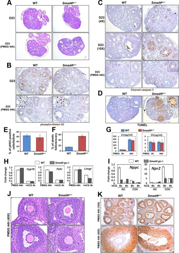
Effects of Smad4 cKO on Follicle Growth, Follicle Atresia, and FSH Target Genes Expression. A, Histology of WT and Smad4gc−/− ovaries at PD23, with or without PMSG treatment. B, Immunohistochemistry results for pHistone H3 (Ser10) indicating GC proliferation in WT and Smad4gc−/− ovaries, with or without PMSG treatment. C and D, Immunohistochemistry results for cleaved caspase 3 (C) and TUNEL assay (D) show an increase in GC apoptosis and follicle atresia in Smad4gc−/− ovaries. E and F, Quantification results of phospho-histone H3 (pHH3) positive cells (E) and TUNEL-positive atretic follicles (F) in WT and Smad4gc−/− ovaries at PD23 with PMSG treatment (44 h). G, Serum estrogen (E2) and P4 concentrations of WT and Smad4gc−/− mice before and after PMSG injections. H, Quantitative RT-PCR results using GCs isolated from WT and Smad4gc−/− ovaries (PMSG 44 h and PMSG 44 h+hCG 4 h) showing the expressions of the FSH target genes Cyp11a1, Fshr, and Lhcgr. I, Quantitative RT-PCR results for the effect of Smad4 depletion on Nppc/Npr2 expression. In WT ovaries before hCG treatment, Nppc was highly expressed in mural GCs and Npr2 was highly expressed in cumulus cells. After hCG treatment, Nppc and Npr2 mRNA levels were down-regulated in mural GCs and cumulus cells, respectively. However, this intricate regulation of Nppc/Npr2 expression was abolished in Smad4gc−/− ovaries. J, Hematoxylin and eosin staining results showing precocious oocyte meiotic resumption in Smad4gc−/− ovaries. K, Immunohistochemistry results for 3β-HSD expression patterns. This enzyme was predominantly expressed by mural GCs of antral follicles in WT ovaries, but was expressed in all GCs and cumulus cells in Smad4gc−/− ovaries.