Figure 5.

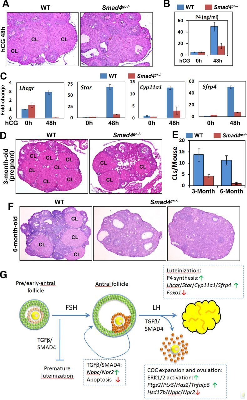
Effects of Smad4 cKO on Luteinization. A, Histology of WT and Smad4gc−/− ovaries at 48 hors after hCG injection. B, Serum P4 concentrations in WT and Smad4gc−/− mice before and after hCG injection. C, Quantitative RT-PCR results. The mRNA expression levels for luteal cell marker genes in WT and Smad4gc−/− ovaries before and after hCG treatments. D–F, Ovarian histology of adult WT and Smad4gc−/− mice (3 and 6 mo old, respectively). At 3 months of age, more CLs were observed in WT ovaries than in Smad4gc−/− ovaries. At 6 months, multiple CLs were observed in WT ovaries but not in Smad4gc−/− ovaries. G, Schematic diagram of SMAD4 functions in GCs during follicle development. The TGF-β family ligands GDF9 and BMP15 secreted by oocytes activate SMADs in nearby granulosa/cumulus cells. SMAD4 depletion in follicles before the antral stage causes premature luteinization of GCs. FSH stimulates follicles to grow beyond the early antral stage and establishes specific gene expression patterns, such as Nppc, Npr2, and 3β-HSD, in mural GCs and cumulus cells together with TGF-β/SMAD4 signaling. However, SMAD4 depletion in follicles beyond the antral stage no longer causes premature luteinization. Rather, SMAD4 is required for preventing follicle atresia at this stage. A TGF-β/SMAD4 signal is also required for LH-induced cumulus expansion, ovulation, and luteinization by inducing ERK1/2 phosphorylation and the expressions of key LH target genes.
