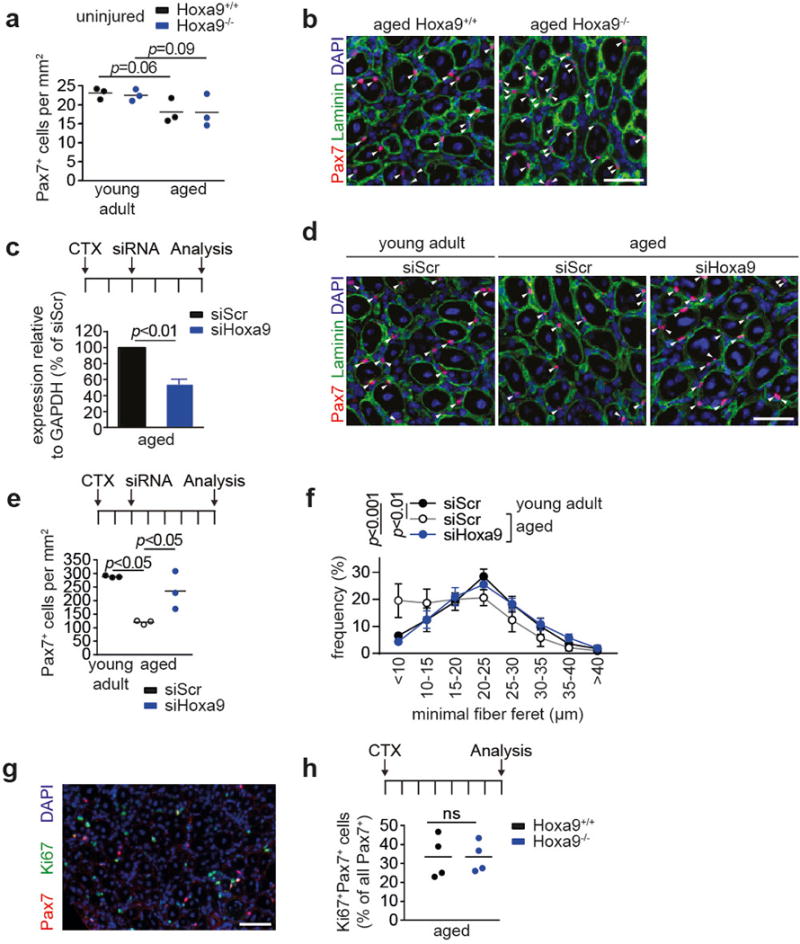
Extended Data Figure 5. Inhibition of Hoxa9 improves muscle regeneration in aging mice.

a, Quantification of Pax7+ cells per area in uninjured tibialis anterior (TA) muscles from young adult and aged Hoxa9+/+ and Hoxa9−/− mice. b, IF staining for Pax7 and Laminin on tibialis anterior (TA) muscles from aged Hoxa9+/+ and Hoxa9−/− mice 7d after Cardiotoxin (CTX) injury. c, qRT-PCR analysis of Hoxa9 expression in SCs isolated from TA muscles injected with a self-delivering Hoxa9 or scrambled siRNA and harvested 5d after muscle injury. d, IF staining for Pax7 and Laminin of injured TA muscles from young adult and aged mice that were injected with a self-delivery siRNA and harvested 7d after muscle injury. Nuclei were counterstained with DAPI (blue). Arrowheads denote Pax7+ cells. e, Quantification of Pax7+ cells from d per area. f, Frequency distribution of minimal fiber feret from d. g, IF staining for Pax7 and Ki67 on TA muscles from aged Hoxa9+/+ and Hoxa9−/− mice 7d after muscle injury. Nuclei were counterstained with DAPI (blue). h, Quantification of proliferating SCs (Ki67+/Pax7+) as depicted in g. Scale bars = 50 μm for b, d, g. Comparisons by two-sided student’s t-test (c, h) or two-way ANOVA (a, e–f). n=3 mice for a; n=3 mice for c; n=3 mice for e–f; n=4 mice for h.
