
Fig. 4. The E57K mutation in Fbln4 has differential effects on elastic versus resistance artery integrity.
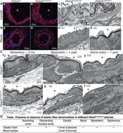
Alexa Fluor 633 hydrazide staining of carotid (A, a and b) and renal arteries (B, a and b) from 3-month-old Fbln4E57K/E57K (b) and WT littermate mice (a). Red indicates elastic fibers, and blue depicts nuclear staining with 4′,6-diamidino-2-phenylindole (DAPI). Mutant vessels are indistinguishable from WT vessels using Alexa Fluor 633 hydrazide staining. Transmission electron micrographs of carotid (A, c and d) and renal arteries (B, c and d) from 3-month-old Fbln4E57K/E57K mice are shown. Micrographs in (d) are higher magnification of the inset demarcated in (c). Note the moth-eaten appearance of the inner two elastic fibers compared to the normal appearance of the outer two elastic fibers in the carotid artery (A, d). (C) Transmission electron micrographs of second-order mesenteric arteries from 3-month-old (a and b) and 1-year-old (c and d) Fbln4E57K/E57K and WT littermate mice in addition to 1-year-old renal arteries. Asterisks indicate vessel lumen. (D) Table summarizing the presence or absence of elastic fiber abnormalities in the different Fbln4E57K/E57K arteries examined.