. 2017 May 3;6:e23966. doi: 10.7554/eLife.23966

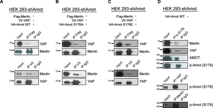
Figure 3. Phosphorylation status of Amot-p130S176 does not impact formation of the Amot/YAP/Merlin complex.

HEK293-shAmot cells were co-transfected with expression plasmids for Flag-Merlin, V5-YAP, and (A) HA-Amot-WT (B) HA-Amot-p130S176A or (C) HA-Amot-p130S176E. Total lysates (input) and Flag or V5 IPs were subjected to immunoblot analysis with anti-YAP and anti-Merlin antibodies as indicated. (D) HEK293-shAmot cells were co-transfected with an expression plasmid for Amot-p130. Total lysates (input) and IPs for phospho-Amot (Ser176), YAP, and Merlin were subjected to immunoblot analysis with indicated antibodies. The blots shown are representative of three independent biological replicates (n = 3).

DOI: http://dx.doi.org/10.7554/eLife.23966.007

Figure 3.

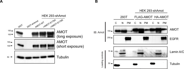
Figure 3—figure supplement 1. Analysis of exogenous Amot expression levels and distribution.

Figure 3—figure supplement 1.

(A) HEK293-shAmot cells were transfected with expression plasmids for Amot-WT, Amot-p130S176A or Amot-p130S176E. Relative levels of Amot expression were assessed by western blotting with anti-Amot antibody and compared to expression of the endogenous protein (293T). Tubulin was used as a loading control. (B) HEK293T or HEK293-shAmot cells that were transfected with expression plasmids for FLAG-Amot-WT or HA-Amot-WT were fractionated in cytoplasmic (C), nuclear (N) or plasma membrane (PM) fractions. Amot was detected by anti-Amot antibody and fractionation was validated by antibodies against EGFR, Lamin A/C and tubulin. The blots shown are representative of three independent experiments (n = 3).
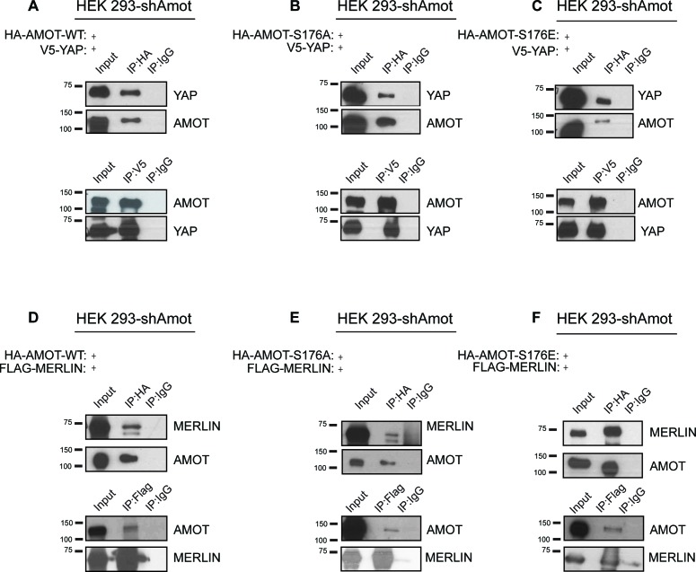
Figure 3—figure supplement 2. Phosphorylation of Amot does not impact formation of YAP/Amot-p130 or Merlin/Amot-p130 complexes.

Figure 3—figure supplement 2.

(A–C) HEK293-shAmot cells were co-transfected with V5-YAP and (A) HA-Amot-WT or (B) HA-Amot-p130S176A or (C) HA-Amot-p130S176E. Total lysates (input) and V5 or HA IPs were subjected to immunoblot analysis with anti-YAP and anti-Amot antibodies as indicated. (D–F) HEK293-shAmot cells were co-transfected with Flag-Merlin and (D) HA-Amot-WT or (E) HA-Amot-p130S176A or (F) HA-Amot-p130S176E. Total lysates (input) and Flag or HA IPs were subjected to immunoblot analysis with anti-Merlin and anti-Amot antibodies as indicated. The blots shown are representative of three independent experiments (n = 3).
Figure 3—figure supplement 3. Phosphorylation of YapS127 does not impact the formation of the Amot/YAP/Merlin complex.

Figure 3—figure supplement 3.

(A) HEK293 cells were co-transfected with expression plasmids for HA-Merlin and His-tagged constitutively active mutant of YAP (His-YapS127A). Total lysates (input) and His or HA IPs were subjected to immunoblot analysis with anti-His and anti-HA antibodies as indicated. (B) HEK293 cells were co-transfected with expression plasmids for Flag-Merlin and YAP. Total lysates (input) and Merlin or phospho-YAP IPs were subjected to immunoblot analysis with anti-phospho-YAP, Merlin and Flag antibodies as indicated. (C) HEK293 cells were co-transfected with expression plasmids for HA-Merlin and Flag-tagged constitutively active mutant of YAP (His-Yap5SA). Total lysates (input) and HA or Flag IPs were subjected to immunoblot analysis with anti-Flag and anti-HA antibodies as indicated. The blots shown are representative of three independent experiments (n = 3).