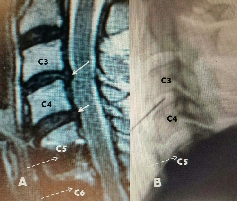
Figure 2. Patient #2: Previous C5-C7 ACDF and plating, new C3-4 disc.
A: T2 sagittal MRI: 2-3 mm C3-4 disc herniation (long white arrow) and smaller C4-5 protrusion (short white arrow). C5 and C6 anterior screws below (dotted white arrows).
B: Intraoperative picture of discectomy probe within C3-4 disc space. The probe can move within the disc space making contact with both superior and inferior endplates. Debridement can be performed with endplate contact.
ACDF: Anterior cervical discectomy and fusion; MRI: Magnetic resonance imaging.

