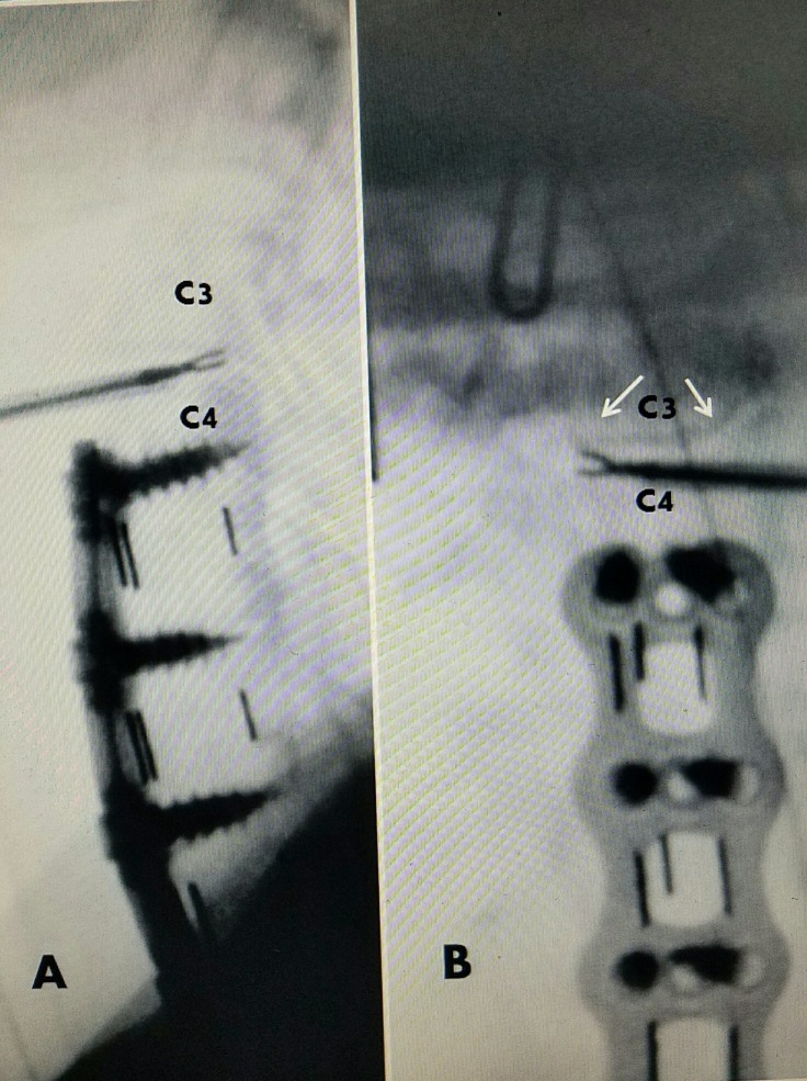
Figure 5. Patient #3: Microtubular manual C3-4 discectomy.
A: Lateral operative picture showing 2.9 mm cannula with 2.5 mm pituitary rongeurs removing posterior-lateral disc at C3-4 above previous fusion and plating C4-C7.
B: AP view showing cannula entering C3-4 disc on patient's left and crossing midline so that pituitary rongeurs is removing disc from right posterior part of the disc space. Uncovertebral joints (two solid white arrows). Cannula enters just medial to UC. The UC prevents pituitary from going too far lateral and confines instrument to disc space.
AP: Anteroposterior.

