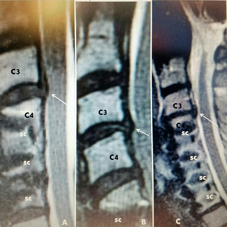
Figure 7. Sagittal MRI scans of three cases.
A: Case #1: previous C4-C6 fusion and plating with screws (sc), small C3-4 disc adjacent level (white arrow).
B: Case #2: C5-C7 fusion and screws (sc) and small herniated disc C3-4 (white arrow).
C: Case #3: Fusion with screws C4-C7 (sc) and slight disc narrowing and subligamentous herniation C3-4 (white arrow).
MRI: Magnetic resonance imaging.

