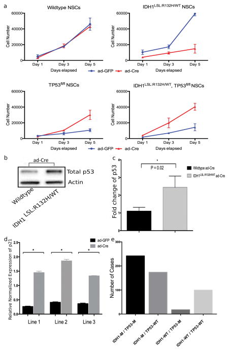
Figure 2. Mutant Idh1-induced cellular arrest is rescued in the presence of Tp53 deletion.
(a) NSCs were infected with ad-GFP or ad-Cre in vitro and expanded. Proliferation was assessed via the CyQUANT Cell Proliferation Assay at Day 1, 3, and 5. Each timepoint contains samples run in triplicate and three independent trials were performed. Figure depicts a representative trial. (b) Western blot analysis of NSCs following ad-Cre infection shows an increase in total p53 in mutant Idh1-expressing NSCs. (c) Quantification of blot intensity of total p53 western blot (unpaired Student t-test, p=0.02, error bars indicate SEM). (d) qPCR for p21 expression was performed on three Idh1LSL:R132H/WT NSC lines treated with either ad-GFP or ad-Cre (normalized to GAPDH) (unpaired Student t-test, * indicates p<0.0001, error bars indicate SEM). (e) Data was extracted from the TCGA database for lower-grade gliomas and samples were categorized based on the mutation status of IDH1 and TP53 (IDH1-M/TP53-M, n=242; IDH1-M/TP53-WT, n=174; IDH1-WT/TP53-M, n=18; IDH1-WT/TP53-WT, n=100; M=mutant, WT=wildtype).

