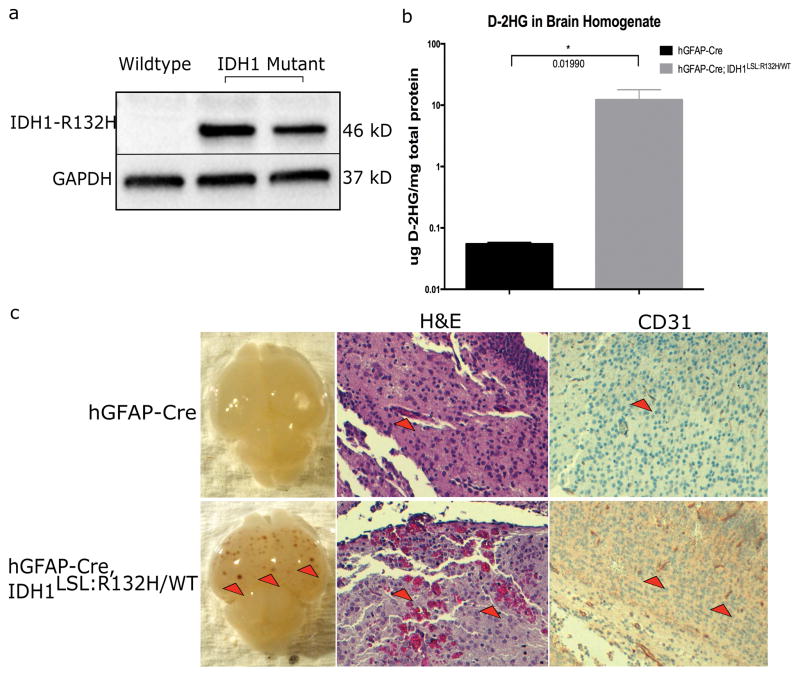
Figure 3. Idh1-R132H is expressed in hGFAP-Cre; Idh1LSL:R132H/WT brains and causes vascular abnormalities.
(a) Western blot analysis using an IDH1-R132H specific antibody on E15.5 brain lysate shows mutant Idh1 is expressed in vivo when under control of the hGFAP-Cre promoter. Blot shows wildtype brain homogenate adjacent to two Idh1-mutant brain homogenates. The production of mutant IDH1 protein was also validated through IDH1-R132H-specific IHC on hGFAP-Cre and hGFAP-Cre; Idh1LSL:R132H/WT brains (data not shown). (b) D-2HG was detected in the brains of hGFAP-Cre; Idh1LSL:R132H/WT animals (n=3 per genotype, unpaired Student t-test, p=0.019, error bars indicate SEM). (c) hGFAP-Cre; Idh1LSL:R132H/WT (bottom row) compared to hGFAP-Cre control animals (top row). Mutant brains (16/17) display areas of hemorrhagic foci (red arrowheads, left panel, bottom row) which were absent from hGFAP-Cre brains (n=20). Red blood cell extravasation was observed (red arrowheads, middle panel, bottom row, 10x) through H&E staining. CD31 staining shows thicker and more robust staining in hGFAP-Cre; Idh1LSL:R132H/WT brains (red arrowheads, right panel, 10x, indicates areas of CD31 positive staining). Phenotype was absent among control animals. Representative images are shown.

