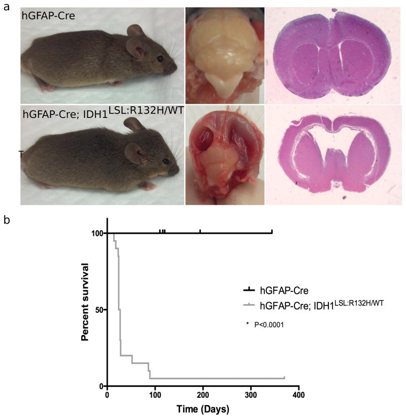
Figure 5. Broad CNS expression of mutant Idh1 induces hydrocephalus.
(a) Comparison of hGFAP-Cre (wildtype (n=9); top row) and hGFAP-Cre; Idh1LSL:R132H/WT (mutant (n=20); bottom row) animals. Gross observation shows head doming and squinted eyes of symptomatic animal (bottom, left). Mutant brains show large, fluid-filled cavities (bottom, middle). H&E staining indicates enlargement of the lateral ventricles and hydrocephalus in mutant brains (bottom, right). (b) Kaplan-Meier survival curve comparing wildtype (hGFAP-Cre) animals (black curve, n=9) and mutant (hGFAP-Cre; Idh1LSL:R132H/WT) animals (grey curve, n=20).

