
Figure 4.
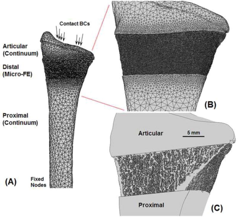
Anterior view of an example multiscale model setup (A) comprising three sections; a continuum articular section, a micro-FE distal section, and a continuum proximal section. Enlarged view showing microstructure details on the surface (B) and through the cross-section (C). Due to partial volume effects in the clinical-resolution images, small edge differences can occur between continuum and micro-FE sections.