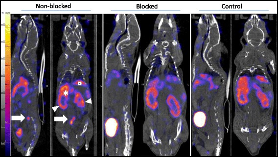
Fig. 3.

Sagittal and coronal views of SPECT/CT images acquired in vivo 90 min after i.v. injection of 111In-tilmanocept from ApoE-KO non-blocked, blocked and control mice. Note the intense focal signals in low abdominal atherosclerotic plaques of ApoE-KO non-blocked mice (white arrows). In contrast, no focal uptake was detected in the aortas of ApoE-KO mice after blocking with excess amount (10 nmol) of non-labelled tilmanocept and in those of control mice. Kidneys (arrowheads) liver (asterisk), spleen (square)
