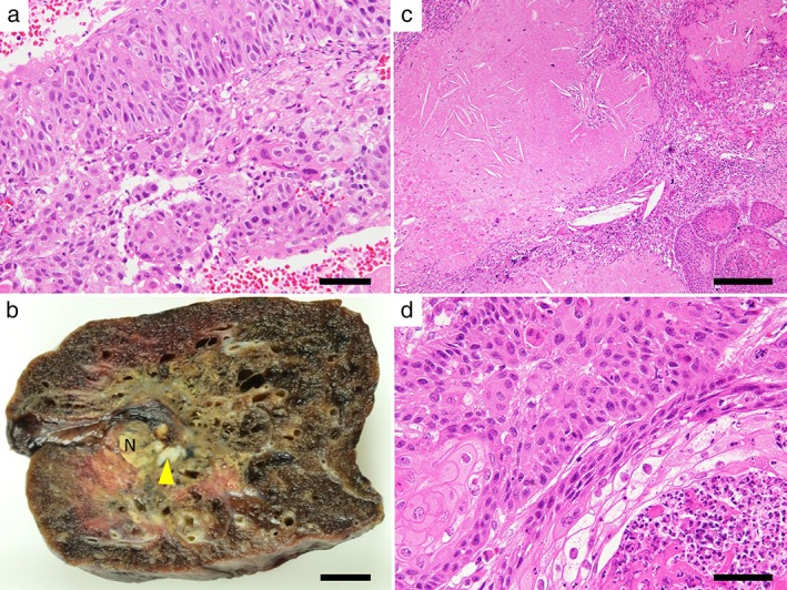
Figure 3.

Macroscopic and microscopic view of the lung cancer. (a) A needle biopsy specimen before chemotherapy showed invasive keratinizing squamous cell carcinoma. Bar = 50 μm. (b) Resected specimen. The tumor had an irregular margin, approximately 1.4 × 1.0 cm, which mostly consisted of collapsed lung parenchyma with fibrosis and necrosis. The yellow granular appearance is a result of tumor necrosis (N). Arrowhead indicates a small focus of residual tumor. Bar = 1 cm. (c) Tumor necrosis with cholesterin clefts and ghost cells. Bar = 200 μm. (d) Residual squamous cell carcinoma with neutrophil inflammation. Bar = 50 μm.
