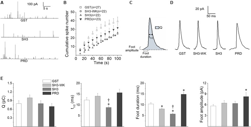
FIGURE 3.

Effects of the cortactin SH3 domain and the N-WASP PRD on exocytosis. Chromaffin cells were injected with 5 μM of GST alone, GST-cortactin SH3 (SH3), a mutated version of GST-cortactin SH3 defective in binding PRD (SH3-WK) or GST-N-WASP PRD (PRD). The exocytosis response evoked by a 10 s pulse of 50 μM DMPP was monitored by amperometry 30–45 min after injections. The amperometric recordings lasted 100 s. (A) Examples of 40 s amperometric traces from cells injected with GST, SH3 or PRD peptides. (B) Cumulative histograms of the number of amperometric spikes from cells injected with GST (white squares), SH3-WK (light-gray circles), SH3 (light-gray triangles) or PRD (dark-gray inverted triangles). Numbers between parentheses indicate the number of cells obtained from at least three different cultures. Notice that both SH3-WK and SH3 significantly reduced the number of spikes between 20 and 80 s. ∗p < 0.05 compared to GST (one-way ANOVA followed by unpaired t-test). (C) Scheme of an amperometric spike with the analyzed parameters: quantal size (Q), half width (t1/2), foot duration and foot amplitude. (D) Examples of amperometric spikes from cells injected with GST, SH3-WK, SH3 or PRD. (E) The graphs show mean values of medians determined for single cells of Q, t1/2, foot duration and foot amplitude of amperometric spikes from cells injected with GST (white bars), SH3-WK (light-gray bars), SH3 (gray bars) or PRD (dark-gray bars). Data are represented as means ± SEM. Numbers of cells for each condition are the same as indicated in (B). ∗p < 0.05 compared with GST; †p < 0.05 compared with SH3-WK (one-way ANOVA followed by unpaired t-test).
