Figure 3.
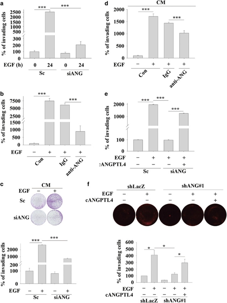
EGF-induced ANGPTL4 enhances HNSCC invasion. The invasive properties of the cells were examined using an invasion assay as described in ‘Materials and methods'. (a) TU183 cells were transfected with 50 nM ANGPTL4 siRNA oligonucleotides (siANG) and scramble siRNA (Sc) by lipofection. Following treatment with 50 ng/ml EGF for 24 h, the invading cells were fixed and stained with crystal violet, solubilized with acetic acid and absorbance (OD, 595 nm) was measured in a microplate reader. Values are the mean±s.e.m. (b) TU183 cells were treated with 10 μg/ml anti-ANGPTL4 antibodies (anti-ANG), 10 μg/ml IgG and 50 ng/ml EGF for 48 h. The invading cells were fixed and stained by crystal violet, solubilized with acetic acid and absorbance (OD, 595 nm) was measured in a microplate reader. Values are the mean±s.e.m. (c and d) TU183 cells were transfected with 50 nM ANGPTL4 siRNA oligonucleotides (siANG) and scramble siRNA (Sc) by lipofection, and CM was collected from EGF-treated or non-treated cells. TU183 cells were then treated with 10 μg/ml anti-ANGPTL4 antibodies (anti-ANG), 10 μg/ml immunoglobulin G (IgG) and CM for 48 h. The invading cells were fixed, stained with crystal violet and examined using a microscope (c, upper panel). Original magnification, × 20. The crystal violet solubilized with acetic acid and absorbance (OD, 595 nm) was measured in a microplate reader (c, lower panel and d). Values are the mean±s.e.m. (e) TU183 cells were transfected with 50 nM ANGPTL4 siRNA oligonucleotides (siANG) and scramble siRNA (Sc) by lipofection, and then treated with 50 ng/ml EGF and 250 ng/ml recombinant C-terminal ANGPTL4 (cANGPTL4) for 48 h. The invading cells were fixed and stained with crystal violet, solubilized with acetic acid and absorbance (OD, 595 nm) was measured in a microplate reader. Values are the mean±s.e.m. (f) The transendothelial invasion of cancer cells was performed as described in ‘Materials and methods'. Endothelial cells were grown to form a monolayer on the bottom of a thick layer of extracellular matrix proteins to mimic intravasation in transendothelial invasion assays. shLacZ and shANG TU183 cells were treated with or without 50 ng/ml EGF and cANGPTL4 for 48 h. The total number of invading cells was quantified by Image J and images were photographed. The percentage of invading cells was determined in three independent experiments. Values are the mean±s.e.m. **P<0.01; ***P<0.005.

