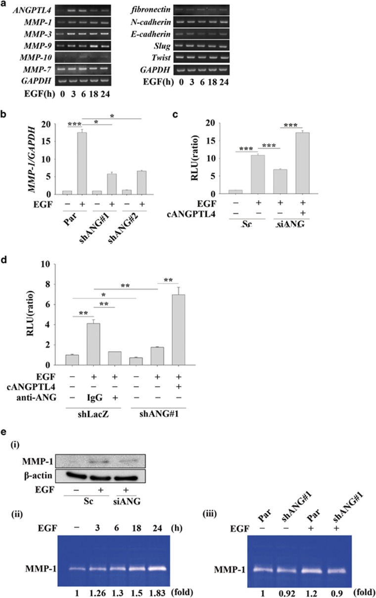
Figure 6.
EGF-induced ANGPTL4 regulates the expression of MMP-1. (a) TU183 cells were treated with 50 ng/ml EGF for the indicated period of time. The expression levels of ANGPTL4, MMP-1, MMP-3, MMP-7, MMP-9, MMP-10, fibronectin, E-cadherin, slug, twist and GAPDH mRNA were analysed by RT-PCR and examined by 2% agarose gel electrophoresis. (b) Parental (Par) and shANGPTL4 (shANG) TU183 cells were treated with 50 ng/ml EGF for 9 h. The expression of MMP-1 mRNA was analysed by real-time quantitative PCR. The relative levels of MMP-1 were normalized to GADPH. Values are the mean±s.e.m. (c) TU183 cells were co-transfected with 0.5 μg MMP-1 promoter and 20 ng renilla luciferase construct, 50 nM ANGPTL4 siRNA oligonucleotides (siANG) and scramble siRNA (Sc) by lipofection. The cells were treated with or without 50 ng/ml EGF and 250 ng/ml cANGPTL4 for 18 h. The firefly and renilla luciferase activities were then determined and normalized. Values are the mean±s.e.m. (d) shLacZ and shANGPTL4 (shANG) TU183 cells were co-transfected with 0.5 μg MMP-1 promoter and 20 ng renilla luciferase construct. Cells were pre-treated with 10 μg/ml of anti-ANGPTL4 antibodies (anti-ANG) and 5 μg/ml IgG for 1 h, and then with 50 ng/ml EGF for 18 h. The firefly and renilla luciferase activities were then determined and normalized. Values are the mean±s.e.m. (e) TU183 cells were transfected with 50 nM ANGPTL4 siRNA oligonucleotides (siANG) and scramble siRNA (Sc) by lipofection, and the cells were treated with or without 50 ng/ml EGF for 18 h. The expression levels of β-actin and MMP-1 were analysed by western blotting (i). TU183 cells were treated with 50 ng/ml EGF for the indicated period of time (ii). Parental (Par) and shANGPTL4 (shANG) TU183 cells were treated with 50 ng/ml EGF for 24 h (iii). The enzymatic activity of MMP-1 in CM was analysed using the casein-zymography assay. *P<0.05; **P<0.01.

