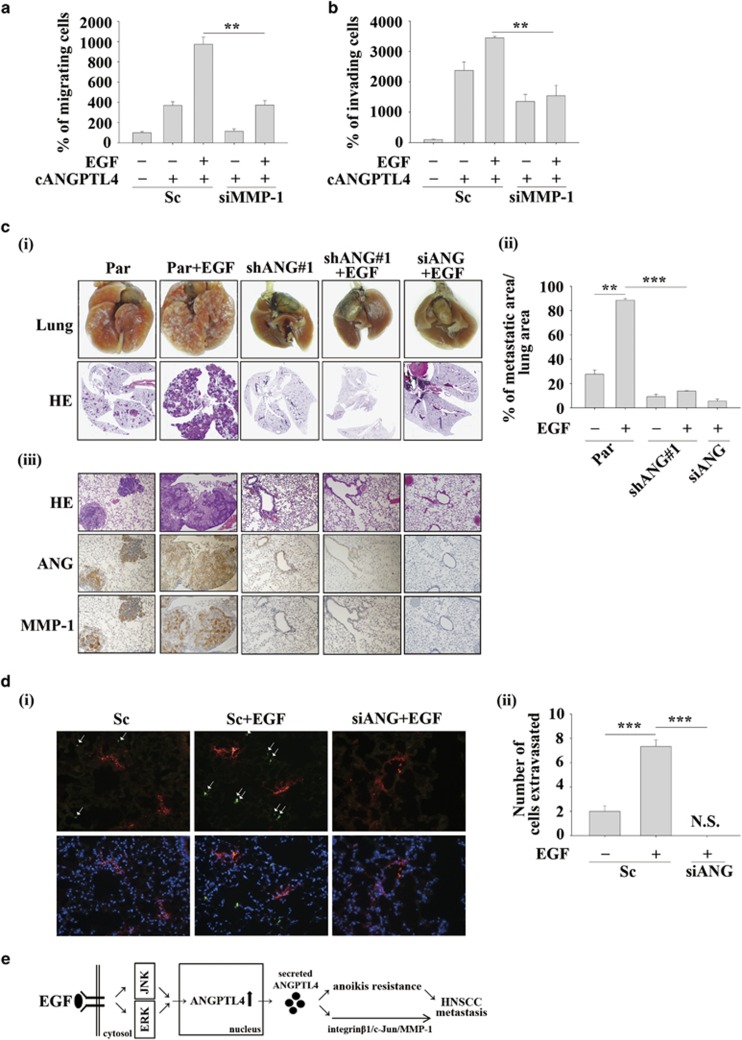
Figure 9.
The EGF/ANGPTL4/MMP-1 axis promotes HNSCC metastasis. (a and b) TU183 cells were transfected with 50 nM MMP-1 siRNA oligonucleotides (siMMP-1) and scramble siRNA (Sc) by lipofection and then treated with 250 ng/ml cANGPTL4 and 50 ng/ml EGF for 24 or 48 h. To determine the percentages of migrating (a) and invading (b) cells, the cells were fixed and stained with crystal violet, solubilized with acetic acid and absorbance (OD, 595 nm) was measured in a microplate reader. Values are the mean±s.e.m. (c) Parental (Par) TU183 cells were transfected with 50 nM ANGPTL4 siRNA oligonucleotides (siANG). Par and shANGPTL4 (shANG) TU183 cells (5x10⁵) were then treated with 50 ng/ml EGF for 3 h and then injected into the tail vein of severe combined immunodeficiency mice. Colonies in the lungs were examined and photographed at 1.5 months. Images of lung and H&E staining (i); the tumour area was analysed using ImageJ (ii); and ANGPTL4 and MMP-1 expression were examined by IHC (iii). n=5 for each groups. (d) Parental (Par) and shANGPTL4 (shANG) TU183 cells were treated with 50 ng/ml EGF for 3 h and then stained with DiO for 30 min. Cells were (5x10⁵ cells/100 μl) injected into the tail vein of severe combined immunodeficiency mice for 24 h and then sacrificed for examining the metastatic tumour cells surrounding the lung tissue as described in ‘Materials and methods'. The penetration of tumour cells was examined using a microscope (i). Original magnification, × 20; arrows indicate the DiO-labelled tumour cells (green); DiI-labelled blood vessels (red); DAPI-labelled nucleus (blue). Tumour cells were quantified by analysing at least four sections and six fields to determine the number of tumour cells that underwent extravasation (ii). n=5 for each groups. Values are the mean±s.e.m. (e) The putative EGF/ANGPTL4/integrin β1/MMP1 pathway regulates HNSCC metastasis. **P<0.01. N.S., non signal (Non DiO-labelled signal was observed).

