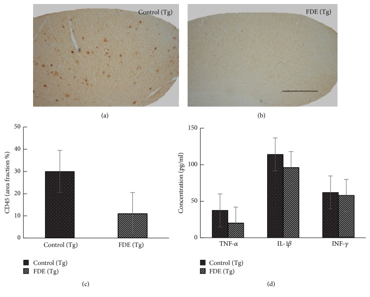
Figure 7.
FDE attenuates inflammation in APP/PS1 transgenic mice. (a–c) Representative images of staining in APP/PS1. No obvious microgliosis was observed in the brain of FDE treatment transgenic mice. (c) Comparison of CD45 area fraction in neocortex among groups. (d) Quantification of IL-1β, IL-6, TNF-α, and IFN-γ in plasma. Denote p < 0.05 or p < 0.01 versus control (Tg) mice, as determined by one-way ANOVA. Scale bar, 1 mm.

