Fig. 1.

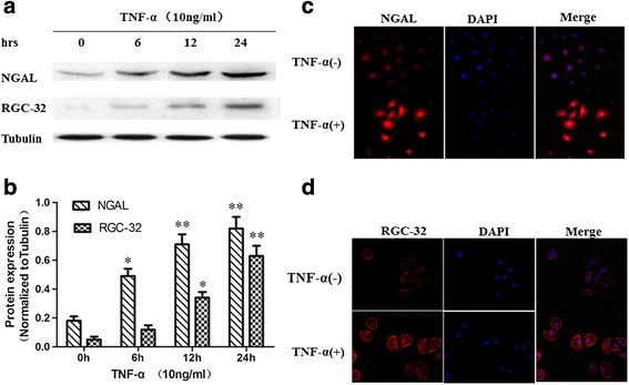
NGAL and RGC-32 expression in NRK-52E cells treated with TNF-α. a – NRK-52E cells were treated with TNF-α (10 ng/ml) and the protein expression of NGAL, RGC-32 and tubulin was detected via western blotting. b – NGAL and RGC-32 expression were normalized to tubulin (n = 5, *p < 0.05, **p < 0.01). c – NGAL localization was analyzed via immunofluorescence staining. NRK-52E cells were treated with TNF-α (10 ng/ml) or untreated for 24 h, and then stained with anti-NGAL primary antibody, followed by incubation with Cy3-conjugated secondary antibody. d – RGC-32 localization was evaluated via immunofluorescence staining. NRK-52E cells were treated with TNF-α (10 ng/ml) or untreated for 24 h, and then stained with anti-RGC-32 primary antibody, followed by incubation with Cy3-conjugated secondary antibody
