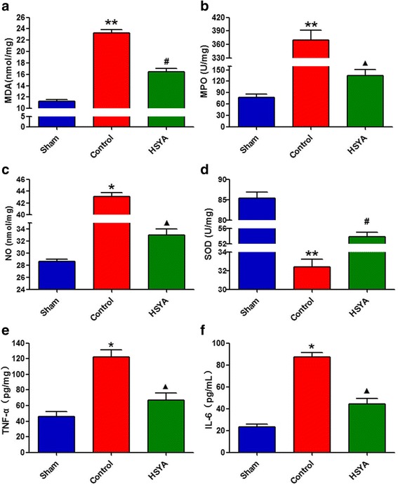
Fig. 3.

Quantification of oxidant stress markers and inflammatory cytokines in spinal cord. a–c Consistent oxidant stress marker parameter results of MDA, MPO, and NO in the control group revealed significantly increase as compared with the sham group 48 h after the lesion. Unanimous as well, these increases were significantly attenuated by HSYA treatment. d Compared with the sham group, the trauma reduced the amount of SOD at 48 h after SCI; however, treatment with HSYA significantly recuperated the amount of SOD. e, f Inflammatory cytokines in spinal cord tissue were detected by ELISA. The levels of TNF-α and IL-6 were significantly increased at 48 h after SCI. On the contrary, HSYA treatment attenuated the SCI-induced increases of these cytokines. (n = 6 *P < 0.05 vs. sham group; **P < 0.01 vs. sham group; ▲ P < 0.05 vs. sham group; P < 0.01 vs. control group; #P < 0.01 vs. control & sham group)
