Fig. 3.

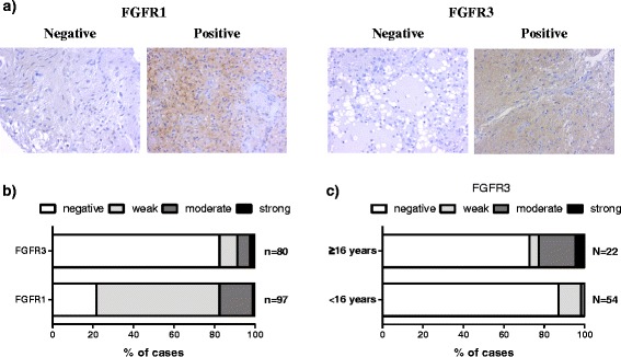
FGFR3 and FGFR1 staining in pilocytic astrocytoma. a Representative immunohistochemical images in pilocytic astrocytoma. b Distribution of immunohistochemistry scores. The majority of samples were negative for FGFR3. c Nearly all of the pilocytic astrocytoma samples showing moderate-to-strong FGFR3 immunostaining were obtained from non-pediatric patients (p < 0.01, Fisher’s exact test). Only newly-diagnosed tumors were included into this analysis
