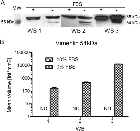
Fig. 7.

Vimentin western blot analysis. HTR-8/SVneo cells were grown in medium with 10 or 0 % FBS for 24 h. Total protein extracts (30 μg) of independent experimental triplicates were separated by SDS-PAGE. Western blot, as previously described, was used to analyze the protein levels of vimentin. a Representative western blot films obtained. MW – molecular weight marker (PageRuler Plus Prestained Protein Ladder, Thermo Scientific). b Densitometric quantifications of expected vimentin band (54 kDa) were made in triplicate to each film using Quantity One software. FBS 10 % ND – not detectable
