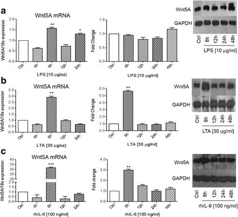
Fig. 1.

Wnt5A expression induced by LPS, LTA and IL-6. SKOV-3 cells were treated with 1 μg/ml LPS (a); 30 μg/ml LTA (b) or 100 ng/ml IL-6 (c) for the indicated times. The left panels show normalized values (means ± SD) from three independent quantitative PCR analyses for Wnt5A expression. Data were normalized related to 18 s RNA as the internal control. The right panels show normalized values (means ± SD) from three independent western blots for Wnt5A. The western blots represent one of three independent experiments. GAPDH levels were used as the internal control. *p ≤ 0.05; **p ≤ 0.01; ***p ≤ 0.001 as compared with the control (Ctrl)
