Fig. 1.

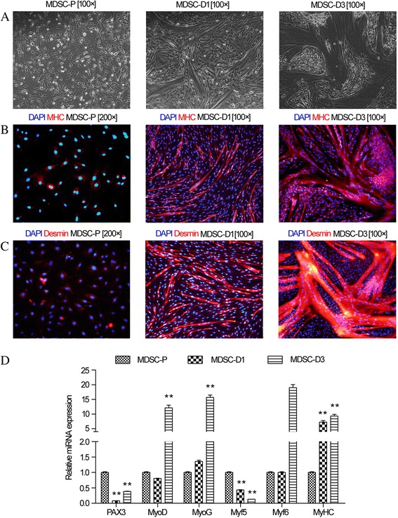
Morphology and immunofluorescence characterization of MDSCs. a Morphology of MDSCs during differentiation for 0, 1, and 3 days (MDSC-P, MDSC-D1, and MDSC-D3, respectively); b Immunofluorescence detection of MHC in MDSCs during differentiation at MDSC-P, MDSC-D1 and MDSC-D3; c Immunofluorescence detection of desmin in MDSCs during differentiation at MDSC-P, MDSC-D1 and MDSC-D3. d Expression of MRF during MDSC differentiation. The RT-qPCR analysis of PAX3, MYOD, MYOG, MYF5, and MYF6 genes in MDSC-D1 and MDSC-D3 compared to MDSC-P. Error bar indicates standard error of mean of triplicate samples. *p < 0.05,**p < 0.01
