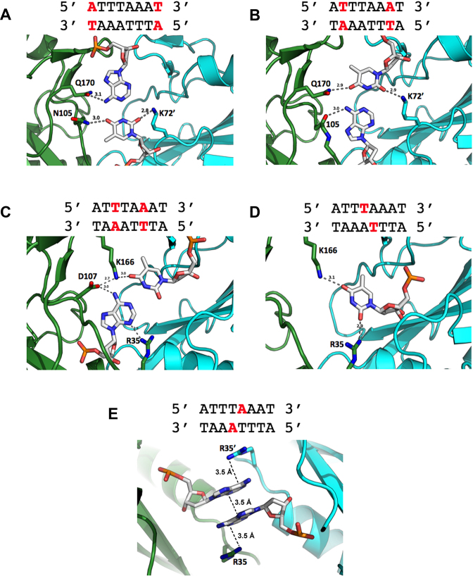
Figure 5.
Atomic contacts made between SwaI and individual bases in its DNA target site. The individual panels show contacts to bases starting at the outermost base pairs (A–C) and progressively working towards the middle of the target site (D and E). For clarity, only contacts to bases in one half-site are shown (except for the adenine bases at the exact center of the target site); the contacts are identical in the two DNA half-sites. The eight bases in each DNA half-site are directly contacted by six amino acid side chains (five from one protein subunit, and one from the opposing subunit). Of those six protein residues, four appear to form bridging contacts to bases at immediately neighboring positions. Two contacts between protein backbone nitrogen atoms (from residues 105 and 107) and atoms on DNA bases (adenine N7 in panels 2 and 3, respectively) are not shown for clarity. Panel e illustrates the position and reversed base-stacking interactions formed between the adenine bases extracted from positions ±1 in the protein-DNA complex. That pair of bases is flanked by the side chains of arginine 35 and 35΄, which respectively form cation–π interactions with each base (while at the same time, also forming contacts to two additional bases). Additional non-specific contacts made between protein side chains and the DNA backbone are also not shown in the figure for clarity.

