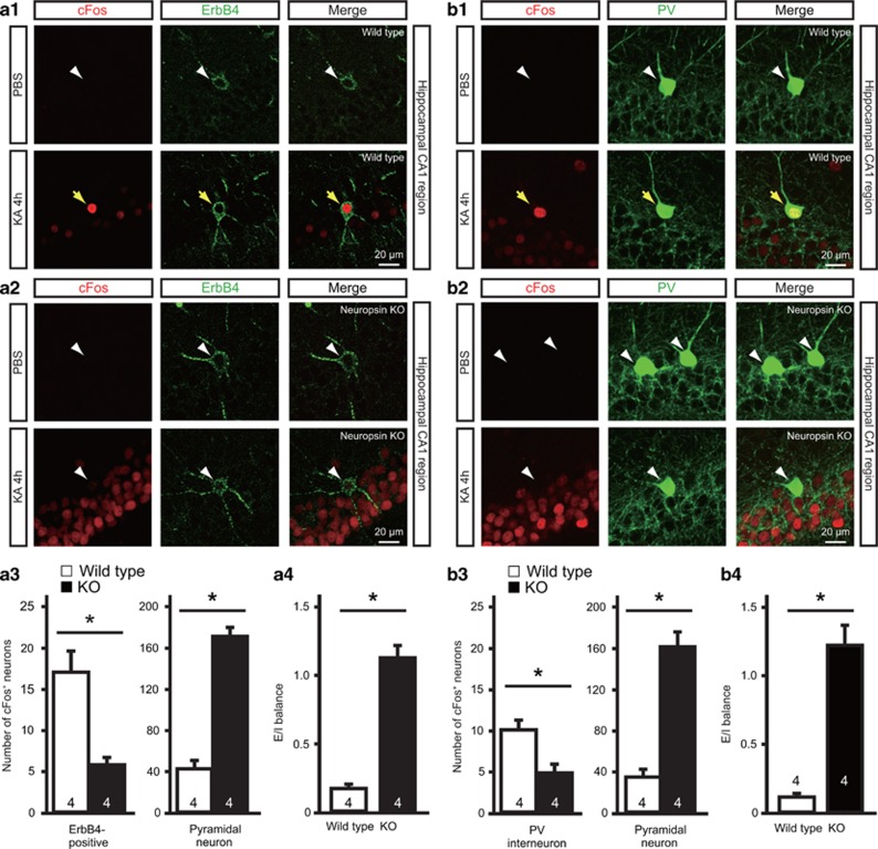
Figure 4.
Neuropsin-KO mice show reduced cFos expression in ErbB4-positive neurons and inhibitory activity and enhanced excitatory activity after seizure. (a1, a2) Expression of cFos in ErbB4-positive and pyramidal neurons of wild-type (a1) or neuropsin-KO (a2) mice 4 h after PBS (upper panel) or KA (lower panel) administration. Fluorescent labeling of the hippocampal CA1 region shows strong cFos immunoreactivity (red) in ErbB4-positive neurons (green) in wild-type mice, but only faint staining in neuropsin-KO mice. Yellow arrows indicate ErbB4-positive neurons expressing cFos. White arrowheads indicate ErbB4-positive neurons that do not express cFos. (a3) Quantitative analysis of cFos immunofluorescent labeling of ErbB4-positive and pyramidal neurons of the wild-type (white bars) and neuropsin-KO (black bars) mice shown in a1 and a2. The number of cFos-expressing ErbB4-positive neurons in neuropsin-KO mice was significantly lower than that in wild-type mice (Mann–Whitney U test, *P=0.0286 vs wild-type); by contrast, the number of cFos-immunoreactive pyramidal neurons from neuropsin-KO mice was significantly higher than that in wild-type mice (Mann–Whitney U test, *P=0.0286 vs wild-type). Data are expressed as the mean and SEM. (a4) E/I balance, defined as the ratio between the fractions of pyramidal neurons and ErbB4-positive neurons expressing cFos, in wild-type (white bar) and neuropsin-KO (black bar) mice. The E/I balance in neuropsin-KO mice was significantly higher than that in wild-type mice (Mann–Whitney U test, *P=0.0286 vs wild-type). (b1, b2) Expression of cFos in parvalbumin (PV)-positive interneurons (arrows) and pyramidal neurons in wild-type (b1) or neuropsin-KO (b2) mice 4 h after PBS (upper panel) or KA (lower panel) administration. Fluorescent labeling of the hippocampal CA1 region shows strong cFos immunoreactivity (red) in parvalbumin-positive interneurons (green) in wild-type mice, but only faint staining in neuropsin-KO mice. Yellow arrows indicate parvalbumin-positive neurons expressing cFos. White arrowheads indicate parvalbumin-positive neurons that do not express cFos. (b3) Quantitative analysis of cFos immunofluorescent labeling in PV-positive interneurons and pyramidal neurons of wild-type (white bars) and neuropsin-KO (black bars) in the mice shown in b1 and b2. The number of cFos-expressing parvalbumin-positive interneurons in neuropsin-KO mice was significantly lower than that in wild-type mice (Mann–Whitney U test, *P=0.0286 vs wild-type); by contrast, the number of cFos-immunoreactive pyramidal neurons in neuropsin-KO mice was significantly higher than that in wild-type mice (Mann–Whitney U test, *P=0.0286 vs wild-type). Data are expressed as the mean and SEM. (b4) E/I balance, defined as the ratio between the fractions of pyramidal neurons and parvalbumin-positive interneurons expressing cFos in wild-type (white bar) and neuropsin-KO (black bar) mice. The E/I balance in neuropsin-KO mice was significantly higher than that in wild-type mice (Mann–Whitney U test, *P=0.0286 vs wild-type). Numbers inside columns indicate n. KA, kainate; KO, knockout; PBS, phosphate buffered saline; SEM, standard error of the mean.

