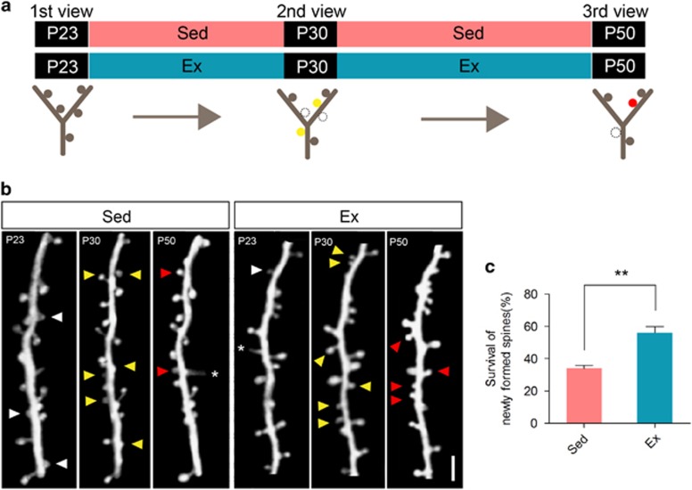
Figure 4.
Reduction of dendritic spine elimination was contributed by enhanced newly formed spine survival. (a) Following first two-photon in vivo imaging of barrel cortex, mice received physical exercise (Ex) or sedentary housing (Sed) from P23 to P50, during which two imaging on the same area were performed. Lower panel: an illustration of one dendritic branch at three time points, showing newly formed spines on P30 (solid yellow circle), which survived (solid red circle) or lost (dash circle) on P50. (b) Two-photon imaging of one dendritic branch on P23, P30 and P50. Eliminated spines (white arrowhead), newly formed spines on P30 (yellow arrowhead), survived newly formed spines on P50 (red arrowhead) and filopodia (asterisk) were shown. Scale bar, 2 μm. (c) Survival rate of newly formed spines on P50. Data were shown as mean±s.e.m. **P<0.01 using Student's t-test. Refer to Supplementary Table 1 for the number of animals and spines in each group.

