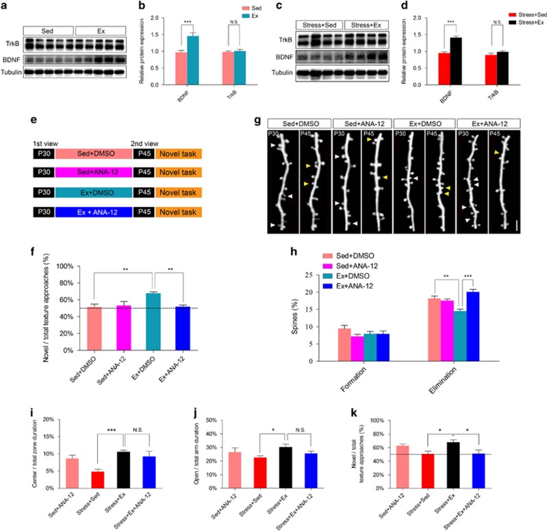
Figure 5.
Physical exercise prevented the loss of spines and improved working memory through BDNF–TrkB pathway. (a and b) Relative protein expression levels of TrkB and BDNF in barrel cortex of naive mouse treated with physical exercise (Ex) or sedentary housing (Sed) from P30 to P45. (c and d) Relative protein expression levels of TrkB and BDNF in barrel cortex of physically constrained mouse treated with physical exercise (Ex) or sedentary housing (Sed) from P30 to P45. (e) Schematic diagram showing two-photon imaging of the same dendritic branch of barrel cortex on P30 and P44, between which mice were sedentary housed (Sed) with DMSO injection or received treadmill exercise (Ex) plus DMSO or ANA-12 injection. (f) Preference for the novel texture in discrimination task. (g) Two-photon images showing eliminated spines (white arrowhead) and newly formed spines (yellow arrowhead) from P30 to P45. (h) Percentage of spine formation and elimination. (i) ANA-12 application did not change duration in the central zone of open filed. (j) ANA-12 injection did not alter the time spent in open arm during elevated plus maze test. (k) ANA-12 injection decreased novel texture recognition ratio of stressed mice even after exercise. Scale bar in g, 2 μm. Data were shown as mean±s.e.m.; *P<0.05, **P<0.01, ***P<0.001 using Student's t-test in b and d, or using one-way analysis of variance (ANOVA) followed by post hoc Tukey test in f–k. N=6–7 animals per group. Refer to the Supplementary Table 1 for the number of spines in each group. BDNF, brain-derived neurotrophic factor; DMSO, dimethyl sulfoxide; NS, no significant difference; TrkB, tyrosine kinase B.

