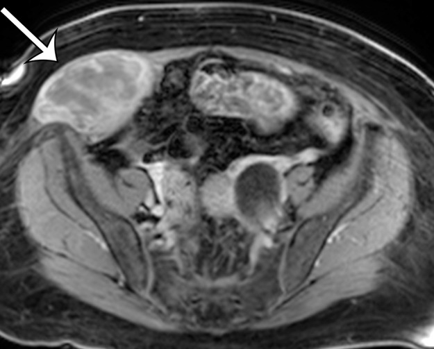
Figure 10d.

Leiomyosarcoma arising in the anterior abdominal wall of a 65-year-old woman with abdominal pain. (a) Axial contrast-enhanced CT image of the upper pelvis shows a homogeneous ovoid mass (arrow) arising within the anterolateral abdominal wall musculature. (b) Axial T1-weighted MR image shows that the mass (arrow) is mildly hyperintense to skeletal muscle. (c) Axial T2-weighted MR image shows that the mass (arrow) is heterogeneous, with curvilinear bands of T2 hyperintensity. (d) Axial contrast-enhanced T1-weighted fat-suppressed MR image shows heterogeneous enhancement of the mass (arrow). (e) Photograph of the gross specimen shows a tan-pink soft-tissue mass with a whorled surface.
