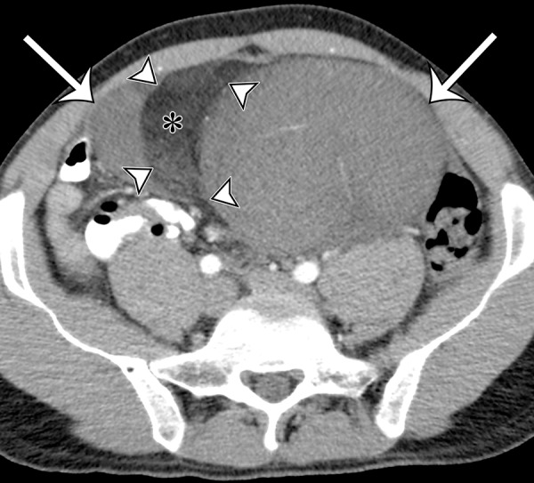
Figure 5a.

Sclerosing type of well-differentiated liposarcoma in a 37-year-old man with severe abdominal pain. (a) Axial contrast-enhanced CT image shows a large multinodular tumor with a fat-attenuation component (*) separated from the soft-tissue enhancing components (arrows) by thin septa (arrowheads). (b) Photograph of the cut surface of the gross specimen shows a similar configuration, with pale-yellow tissue correlating with the fat component (*) shown on a, and with pink-yellow tissue correlating with the soft-tissue components (arrow); the components are separated by fibrous septa (arrowheads). (c) Photomicrograph of the sclerosing type of well-differentiated liposarcoma shows a few adipocytes and nonlipomatous cells in a collagenous matrix. (Hematoxylin-eosin [H-E] stain; original magnification, ×40.)
