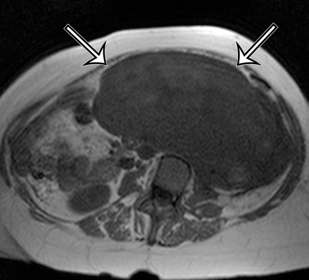
Figure 7a.

Incidentally identified intra-abdominal leiomyosarcoma in a 55-year-old woman. (a–c) Axial T1-weighted (a), T2-weighted (b), and gadolinium-enhanced T1-weighted fat-suppressed (c) MR images show a large relatively homogeneous, non–fat-containing solid intra-abdominal mass that is isointense to skeletal muscle on T1-weighted images (arrows on a), heterogeneously T2 hyperintense (arrows on b), and heterogeneously enhanced (arrows on c). (d) Photomicrograph of leiomyosarcoma shows elongated atypical spindle cells with blunt-ended nuclei with mitotic activity. (H-E stain; original magnification, ×80.)
