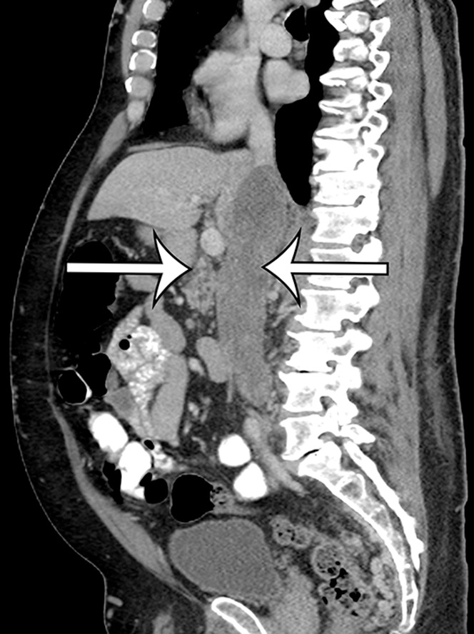
Figure 8b.

Intraluminal and extraluminal leiomyosarcoma in the IVC of a 46-year-old man who presented with a 5-month history of progressive leg swelling. Coronal (a) and sagittal (b) contrast-enhanced CT images show a heterogeneously enhancing mass expanding the IVC (straight arrows), with a small round extraluminal component inferiorly (curved arrow on a).
