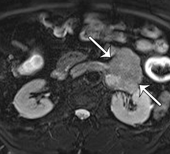
Figure 9b.

Leiomyosarcoma arising in the left renal vein in a 59-year-old man with progressive left flank pain. (a, b) Axial T1-weighted (a) and T2-weighted (b) MR images show a homogeneous mass intimately associated with the left renal vein, with an extraluminal component that is isointense to skeletal muscle on T1-weighted images (arrows on a) and mildly hyperintense to skeletal muscle on T2-weighted images (arrows on b). (c) Coronal gadolinium-enhanced T1-weighted fat-suppressed MR image shows enhancement of the central vein component (straight arrows) and the intraluminal tumor extending into the infrahepatic IVC (curved arrow). (d) Photograph of the gross specimen shows the intraluminal component adherent to the wall of the left renal vein (straight arrows) and the contiguous smooth gray-tan tumor thrombus extending beyond the resected renal vein margin (curved arrows); the tumor thrombus was milked from the IVC.
