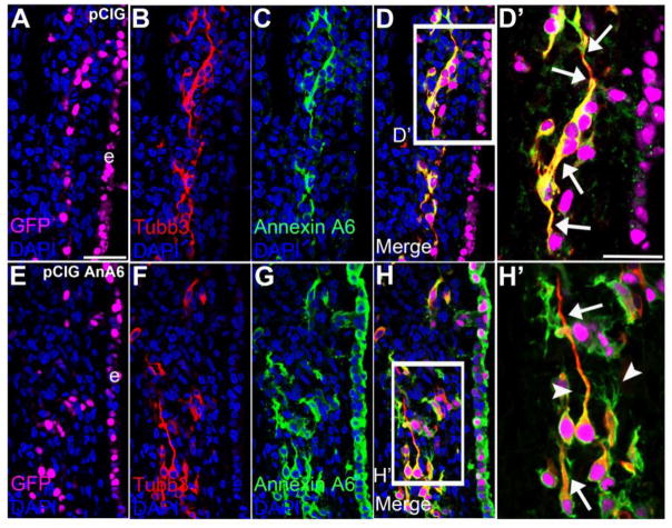
Figure 10. Annexin A6 overexpression in sensory placode cells does not affect placode cell ingression, differentiation, and position within the ganglionic anlage.
Representative transverse section of the forming trigeminal and ganglion at HH16 after electroporation of pCIG control (A–D′) or pCIG-Annexin A6 (E–H′, pCIG AnA6) expression construct at HH10. Trigeminal neurons electroporated with the control pCIG (A, GFP, false-colored in purple) exhibit a bipolar morphology as seen by Tubb3 immunostaining (B, D, D′, arrows), while those overexpressing Annexin A6 (E, GFP, false-colored in purple) also show two processes through Tubb3 (F, H, H′, arrows). Some neurons containing pCIG-Annexin A6 (G–H′) possess extra protrusions emanating from their cell bodies and the membranes of their bipolar processes (H′, arrowhead). DAPI (blue) labels cell nuclei. e, ectoderm. Scale bar, 50μm (A–H) and 25μm (D′, H′).

