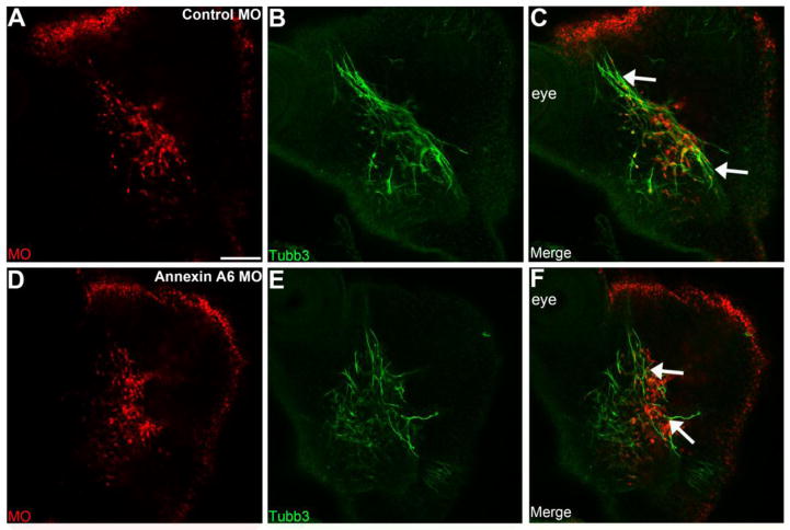
Figure 5. Annexin A6 knockdown in sensory placode cells alters the morphology of the trigeminal ganglion.
Representative lateral views (optical section) of the forming trigeminal ganglion in an HH16 chick head after electroporation of a 5 bp mismatch control Annexin A6 (A-C) or Annexin A6 (D-F) MO at HH10. Trigeminal ganglia electroporated with the control MO exhibit a condensed, organized morphology, with forming nerve bundles, as seen by Tubb3 immunostaining (C, arrows), while those electroporated with the Annexin A6 MO possess a disorganized and dispersed morphology, with neurons present throughout the embryo head (F, arrows). Scale bar, 50 μm (A) applies to all images.

