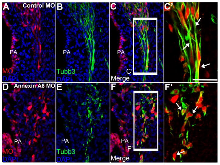
Figure 6. Annexin A6-depleted neurons lack processes to innervate their designated targets.
Representative transverse section of the forming trigeminal ganglion at HH20 after electroporation of a 5 bp mismatch control Annexin A6 (A–C′) or Annexin A6 (D–F′) MO at HH10. Maxillary and mandibular branches of the trigeminal neurons electroporated with the control MO possess neuronal processes, as seen by Tubb3 immunostaining (B–C′), and form nerves with neighboring neurons to innervate the pharyngeal arch (C′, arrows). Trigeminal neurons possessing Annexin A6 MO show few to no processes (E–F′) and, consequently, do not form nerves to innervate the pharyngeal arches (F′, arrows). DAPI (blue) labels cell nuclei. PA, pharyngeal arch. Scale bar, 50μm (A–F) and 25μm (C′, F′).

