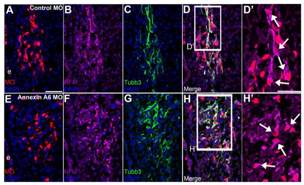
Figure 8. Annexin A6-depleted trigeminal neurons express the neuronal maturation marker NFM.
Representative transverse section of the forming trigeminal ganglion at HH16 after electroporation of a 5 bp mismatch control Annexin A6 (A-D’) or Annexin A6 (E-H’) MO at HH10. Control MO-treated placode cell-derived neurons (A, D, D’) that are Tubb3 immunoreactive (C, D) express NFM (B, D, D’, arrows). Annexin MO-containing cells (E, H, H’), which possess few to no protrusions as evidenced by Tubb3 immunostaining (G, H), also express NFM (F, H, H’, arrows). DAPI (blue) labels cell nuclei. e, ectoderm. Scale bar, 50 μm (A-H) and 25 μm (D’, H’).

