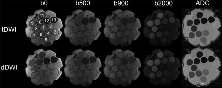
Figure 2.
Representative diffusion-weighted imaging (DWI) images and ADC maps using tDWI and dDWI of a dedicated NIST diffusion phantom (containing 13 vials) at 1.5 T (see Table 1 for sequence parameters). Similar ADC quantification and image quality was observed between tDWI and dDWI.

