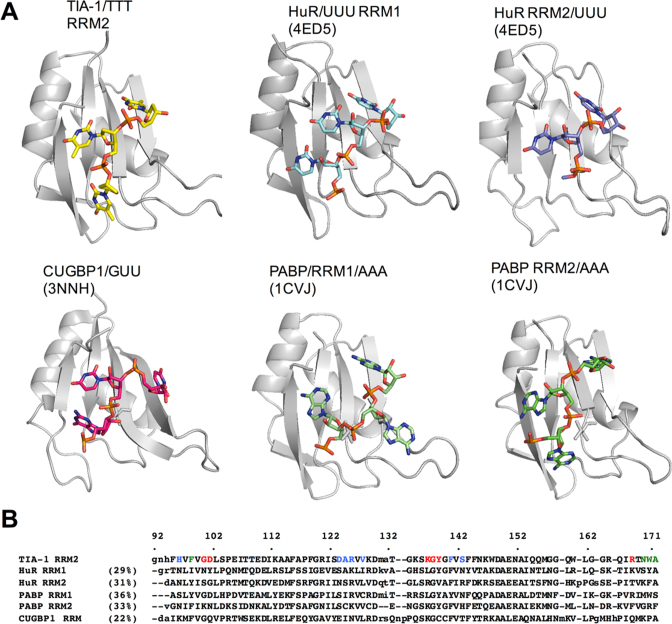
Figure 6.
Comparison of the TIA-1 RRM2 structure with other RRM/RNA interactions. (A) Structural representations of RRM/oligonucleotide interactions determined for HuR (PDB ID: 4ED5), CUGBP1 (PDB ID: 3NNH) and PCBP (PDB ID: 1CVJ) shown in the same orientation as that for TIA-1 RRM2/TTT. (B) Structure-based sequence alignment of the depicted RRMs. The TIA-1 RRM2 amino acids engaged in interactions with thymine are colored in red (T1), green (T2) and blue (T3).

