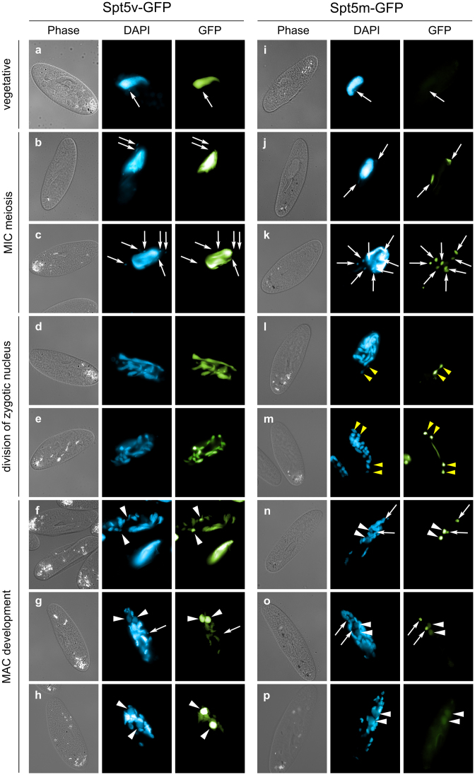
Figure 2.
Localization of Spt5-GFP fusion proteins. For both SPT5v-GFP and SPT5m-GFP transgenes, representative images illustrate different developmental stages. Panels a and i show vegetative cells. Successive stages of autogamy are shown in the following panels: panels b and j—meiotic crescent stage; panels c and k—cells with eight haploid nuclei resulting from meiosis II (only six nuclei are visible in panel C); panels d, e, l and m—fragmentation of old MAC; panel L—first division of the zygotic nucleus; panel M—second division of the zygotic nucleus; panels f and n—early MAC development; panels g, h, o and p—late MAC development. In all panels, white arrows point at MICs (some were omitted when MICs were not clearly distinguishable by DAPI staining), white arrowheads indicate new MACs. Yellow arrowheads in panels L and M point to division products of the zygotic nucleus. The Spt5v-GFP fusion (left side) localizes to old, then new MACs. The Spt5m-GFP (right side) localizes to meiotic MICs, the zygotic nucleus as well as products of division of the zygotic nucleus.

