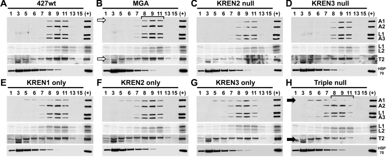
Figure 3.
Western and adenylation analyses of glycerol gradient fractionated editosomes. Gradient fractions from 427wt (A), MGA (B), KREN2 null (C), KREN3 null (D), KREN1 only (E), KREN2 only (F), KREN3 only (G) and triple null (H) were analysed using antibodies recognizing editosome proteins KREPA1, KREPA2, KREL1 and KREPA3 (first panels), by adenylation of ligases KREL1 and KREL2 (second panels), using antibody against KRET2 (third panels), or using antibody against mitochondrial HSP70 as a non-editosome control. Typical ∼20S editosome peak signal is centered on fraction 9, as highlighted by the bracket in the MGA control. In KREN1 only, KREN2 only, KREN3 only, and Triple null cells the sedimentation of KREPA1 is notably shifted toward upper part of the gradient (i.e. smaller in size) relative to 427wt and MGA controls, as is KRET2 in Triple null cells (indicated by solid arrows in Triple null compared to open arrows in MGA). Bracket in Triple null cells highlights difference in the ∼20S region compared to MGA, A control sample of ∼20S fraction from purified mitochondria (+) is included in each analysis.

