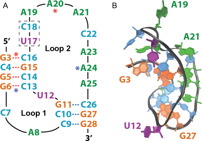
Figure 1.
Luteovirus pseudoknot structure. (A) Sequence and secondary structure, with Stem 1 (S1) beginning at the 5΄ terminus, Stem 2 (S2) ending at the 3΄ terminus, and the locations of the C17U/A18C mutations noted by the dashed gray box. Red and blue asterisks denote Loop 2 (L2) residues that form base-triplets with S1 base pairs. (B) Three-dimensional representation of the crystal structure (PDB ID: 2A43) with selected residues labeled to aid in visual orientation.

