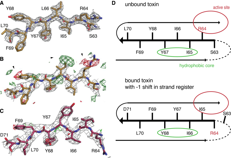
Figure 5.
Sliding of VcHigB2 strand β3. (A) Model of the strand β3 from the Nb6-VcHigB2 structure and the corresponding 2mFo-DFc map contoured at 1 σ (resolution 1.85 Å). (B) Positive (green) and negative (red) peaks in the mFo-DFc map contoured at 3 σ suggest a different tracing of the sequence of strand β3 in the VcHigBA2 complex. (C) 2mFo-DFc and mFo-DFc maps (contoured at 1 σ and 3 σ, respectively; resolution 3 Å) for the final model of strand β3 in the VcHigBA2 complex. (D) Schematic figure displaying strand β3 in both conformations. Unstructured β2-β3 loop is shown as dashed line, locations of the active site and solvent-inaccessible hydrophobic core are circled. Slided conformation (bottom) does not perturb the hydrophobic packing due to a sequence repeat.

