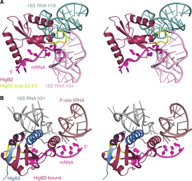
Figure 6.
Model of the VcHigB2 toxin bound to the ribosome. The model was obtained by the superposition of the VcHigB2 toxin on EcRelE in the EcRelE–ribosome complex (PDB ID: 4V7J). (A) VcHigB2 positively charged β2-β3 loop (highlighted in yellow) interacts with helies 18 and 34 from the 16S RNA. The conformation of the β2-β3 loop was optimized using ModLoop web server (61). (B) VcHigB2 toxin with bound VcHigA23-33 peptide placed in the VcHigB2 binding site of ribosome using the EcRelE–ribosome complex (PDB ID: 4V7J) as a guide. No obvious clashes are observed between the ribosome or the mRNA substrate and VcHigB2 or VcHigA23-33.

