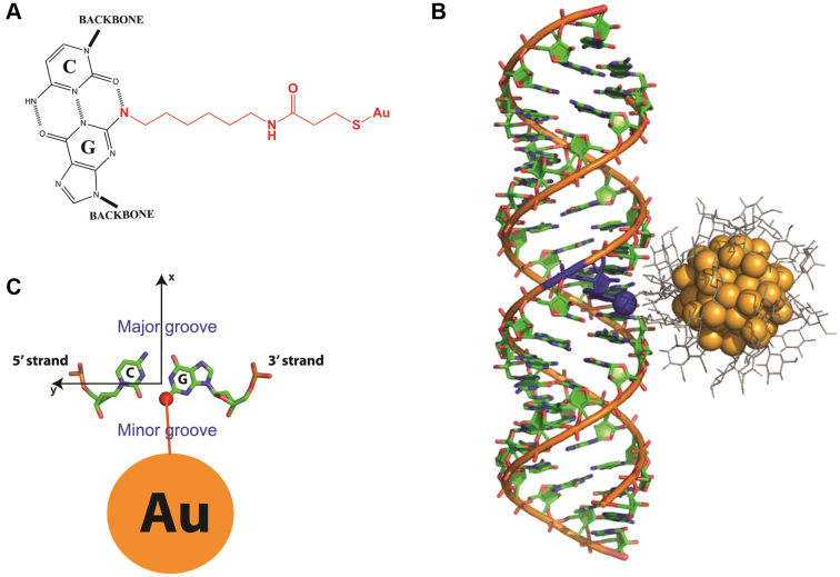
Figure 1.
The position of Au nanocrystals internally labeled to RNA helix. (A) The internally labeled nanocrystals are attached to N2-amino-modifed Gs through SPDP (N-Succinimidyl 3-[2-pyridyldithio]-propionate) linkers. (B) The Au nanocrystal shown in the plane of its attachment to G (seen from top). (C) The XSI-determined model of a thio-glucose (grey) passivated Au nanocrystal (orange) attached to an RNA helix. The attachment is to N2 (blue sphere) of a G residue (blue) as shown in part A, which positions the Au nanocrystals in the minor groove of the RNA helix.

