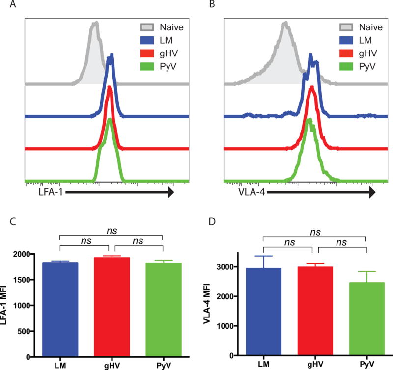
Figure 5. Donor-specific CD8+ T cell surface expression of LFA-1 and VLA-4 does not explain altered susceptibility profiles to CoB/integrin therapy.

Naïve C57BL/6 mice were adoptively transferred 104 OT-I T cells, infected with OVA-expressing LM, gHV or PyV, and peripheral blood lymphocytes analyzed for surface expression of LFA-1 and VLA-4 30 days after infection. (A & B) Representative surface expression of LFA-1 (A) and VLA-4 (B) on CD8+Thy1.1+ memory T cells compared to naïve T cells. (C & D) Summary data of LFA-1 (C) and VLA-4 (D) MFI on CD8+Thy1.1+ T cells (n = 8–10 per group). Summary data represent mean (SE).
