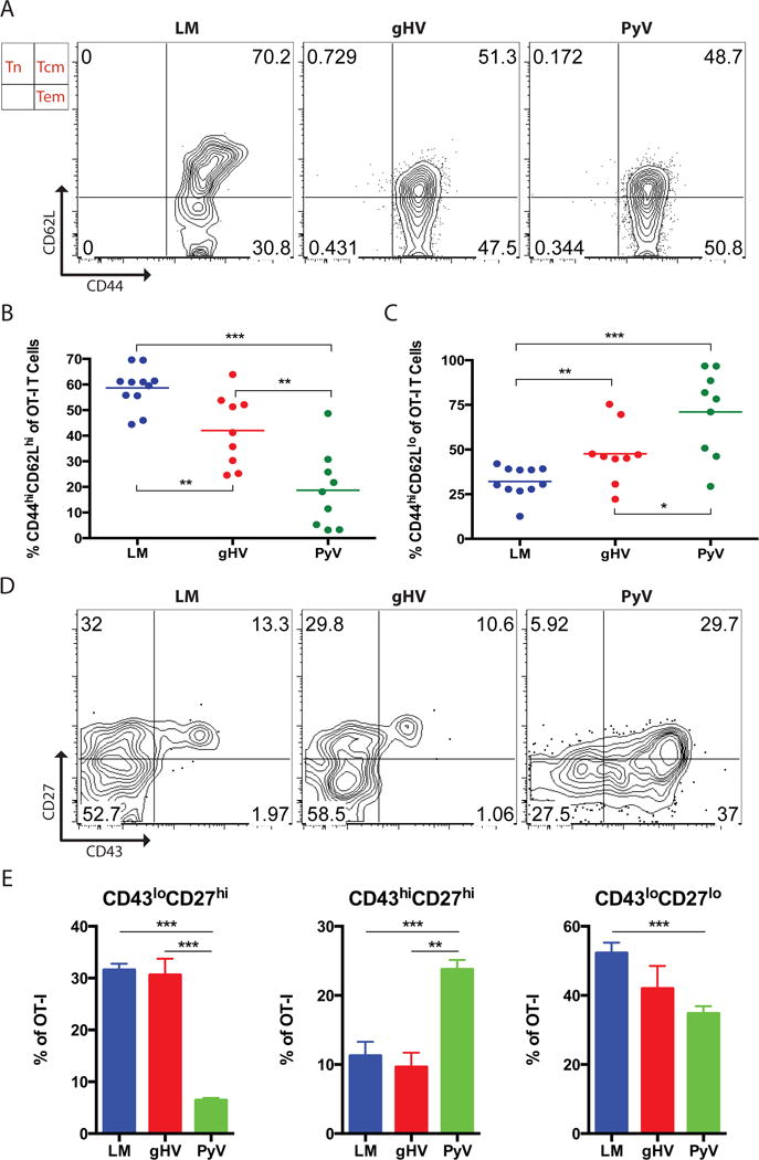
Figure 6. Donor-specific CD8+ memory T cells most susceptible to CoB/integrin blockade exhibit a TCM phenotype and are CD43loCD27hi.

Naive C57BL/6 mice were adoptively transferred 104 OT-I T cells and then infected with OVA-expressing LM, gHV or PyV. Peripheral blood lymphocytes were then characterized 30 days after infection. (A) Analysis of CD44 and CD62L expression on antigen-specific memory T cells. Flow cytometry plots are representative and gated on CD8+Thy1.1+ T cells. (B & C) Summary data of the frequency of CD8+ antigen-specific CD44hiCD62Lhi TCM (B) and CD44hiCD62Llo TEM (C) T cells (n = 8–10 per group). (D) Analysis of CD43 and CD27 expression on antigen-specific memory T cells. Flow cytometry plots are representative and gated on CD8+Thy1.1+ T cells. (E) Summary data of the frequency of CD43loCD27hi, CD43hiCD27hi, and CD43loCD27lo T cells (n = 8–10 per group). Summary data represent mean (SE). * p < 0.05, ** p < 0.01, *** p < 0.001.
