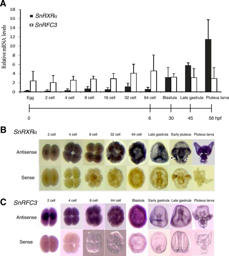
Fig. 7.

Temporal and spatial mRNA expression levels of RXRα and RFC3 in sea urchin embryonic development. A, Temporal expression levels of S. nudus RXRα (SnRXRα) and RFC3 (SnRFC3) mRNA using real-time PCR. The relative SnRXRα and RFC3 mRNA levels were normalized by ubiquitin values and expressed as mean ± sd (n = 4 maternally independent individuals). B and C, Spatial distribution of SnRXRα or SnRFC3 transcripts during normal embryonic development analyzed by whole-mount in situ hybridization. The embryos were hybridized with an antisense or sense probe as indicated. The 32-cell, 64-cell, and blastula stages are animal-pole views. The gastrula- and pluteus-stage embryos are in lateral view with the animal pole at the top. Dashed-line squares and arrowheads indicate the gut and nonspecific signals, respectively. hpf, Hours post-fertilization.
