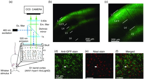
Fig. 1.
Experimental setup and histological validation of ArcLight expression. (a) The experimental setup for ArcLight imaging. (b) Confocal image of the characteristic spread of ArcLight in the S1 barrel cortex (see Sec. 2). Fluorescence (green) from ArcLight excited with 465-nm LED. Layers based on characteristic depths are outlined in white, cross validated with Nissl stain. (c) Confocal image of ArcLight expression. The ArcLight expression can be clearly seen across layer and layer 5. (d) Confocal image of ArcLight expression in cortical region cryosectioned and stained using an anti-GFP polyclonal antibody. Fluorescence is clearly expressing in the neural membranes. An example cell is highlighted with the white arrow. (e) Same section as (d) stained with Nissl (red) for identification of neural cell bodies. (f) Merged image from (d) and (e) shows fluorescent expression in membranes surrounding Nissl (red) stained neural somas. Expression appears to be targeted to somatic, dendritic, and axonal neural membranes.

